


- HYGEIA
- Services


Neurosurgery and Interventional Neuroradiology
The Department treats all brain and spinal cord conditions (e.g. tumors, hematomas, abscesses, vascular malformations, hydrocephaly, etc) that require surgical treatment with open craniotomy-trephination, or with endovascular surgery (known as embolization). It specializes in CNS vascular malformations.The Neurosurgery & Interventional Neuroradiology Department is divided into two units:
a) Interventional Neuroradiology
b) Neurosurgery
These two units treat all brain and spinal cord conditions (e.g. tumors, hematomas, abscesses, vascular malformations, hydrocephaly, etc) that require surgical treatment with open craniotomy-trephination, or with endovascular surgery, better known as embolization.
Although the Department covers all Central Nervous System (CNS) surgical conditions (i.e. brain and spinal cord), the Department physicians also specialize in treating CNS vascular lesions, performing a large number of such procedures every year. As a result, the Department is among the largest and most experienced Vascular Neurosurgery departments worldwide.
Interventional Neuroradiology is a new and rapidly developing medical subspecialty which uses special techniques and the latest materials to treat vascular conditions of the Central Nervous Systems (CNS) without open surgery, by using endovascular approaches (i.e. through blood vessels). Due to the advancement of technology with state-of-the-art imaging techniques and advanced materials, the interventional neuroradiology applications have been expanding and covering all the more conditions and, consequently, patients.The Department treats patients with vascular conditions of the brain and spinal cord, such as brain aneurysms, brain and spinal cord arteriovenous malformations, CNS vascularized lesions and more. It is the largest such department in Greece, handling more than 300 cases annually, a number comparable to those of the largest centers abroad. The fact that its physicians participate in Greek and international conventions as well as have articles published in renowned scientific journals reflects the Department’s multifaceted activities.
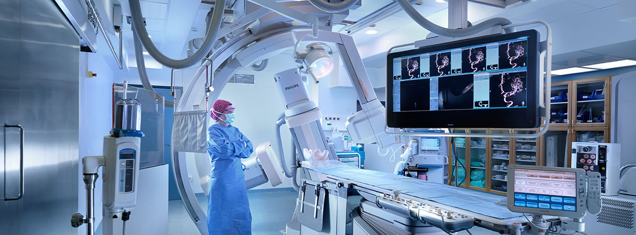
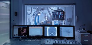
The operating room has been renovated recently, and is equipped with the latest technology (biplane digital angiography device with 3D recomposition capacity) and staffed by qualified doctors, nurses and technicians.
It is on call 24/7 and can also treat emergencies, in partnership with other Hospital departments (CT & MRI Department, ICU).
The Interventional Neuroradiology Department offers the following services:
• Brain vascular malformation embolization
• Brain aneurysm embolization
• CNS neoplasm embolization
• Carotid angioplasty
• Balloon test occlusion (BTO)
• Digital subtraction angiography of the brain
• Digital angiography of the neck
• Digital angiography of the spinal cord
• Digital angiography of the aortic arch/carotidsConditions/Procedures
The most commonly encountered conditions in Interventional Neuroradiology are outlined below, along with the respective procedures used to treat them. Diagnostic access for these conditions is established using the latest imaging techniques (CT or MRI scans), which can be performed within the Hospital, or with intra-arterial digital angiography of the brain and spinal cord, which is considered the most accurate diagnostic method to date. Digital angiography is performed under local anesthesia, through femoral artery catheterization and subsequent investigation of the carotid and spinal arteries.
Brain Aneurysms
An aneurysm is a localized dilation of a blood vessel in the brain, usually occurring at branch points, and has a weak and thin wall. The annual rate for aneurysm rupture is 4%. When an aneurysm ruptures, a life-threatening condition develops, known as subarachnoid hemorrhage. In this case, the aneurysm must be occluded to prevent recurrence of hemorrhage in the future, which may even prove life-threatening.
Two methods are used to treat aneurysms:
A. Craniotomy (open surgery) – a tiny clip is placed across the neck of the aneurysm (see Neurosurgery Department).B. Embolization – platinum microcoils are placed in the aneurysm. The specialist uses a microcatheter to penetrate the aneurysm and introduce the microcoils, packing it to the point where no blood can circulate within the aneurysm. The microcatheters are inserted in the human body via femoral artery catheterization, avoiding opening the skull.

Image 1:
Left. A microcatheter is inserted in the femoral artery through the gluteus area (thick arrow below) and enters the brain vessels (thin arrow above) through the arterial system (aorta, carotid).
Right. Visual representation of embolization. The end of the microcatheter is inserted in the aneurysm (arrow) and platinum coils are introduced to the aneurysm through it.
Image 2:
On the left – aneurysm before embolization (arrow).
On the right – the arrow points to the position of the embolized (i.e. occluded) aneurysm.Brain & Spinal Cord Arteriovenous Malformations
Arteriovenous malformations (AVM) are abnormal connections between arteries and veins in the brain. These malformations are congenital (i.e. they develop when the fetus is growing within the mother’s uterus) and, therefore, may cause problems at any age (infants, babies, children, adolescents and adults). In an angiography, they appear as a “tangle” or “mesh” of malformed blood vessels with increased blood flow within them. These brain malformations lead to symptoms such as headaches, epileptic seizures or even cerebral hemorrhage. The respective spinal cord malformations manifest neurological symptoms, such as weakness in the upper or lower limbs and loss of bladder control. AVM rupture is a serious condition that can cause severe symptoms due to the development of CNS hemorrhaging. The annual rate for brain AVM rupture is 2-4%.

Image 3:
Left. AVM (specifically, this is known as vein of Galen malformation) in a 2.5-month-old baby. A tangle of arteries (small horizontal arrow) pumps arterial blood into a large dilated vein (large arrow). The normal brain vessels are hardly visible (double arrows).
Right. Total occlusion of malformation following embolization. The brain vessels are fully enhanced (double arrows).Interventional neuroradiology plays a leading role in the treatment of brain and spinal cord AVMs in children and adults. Using state-of-the-art angiography devices and special advanced microcatheters, it is possible to perform catheterization of the arteries that supply blood to the malformation, and occlude it using special synthetic materials. This technique can be performed on its own or coupled with other treatments (surgical approach, Gamma knife), depending on the size, location and clinical manifestation, to achieve the best possible result, which is full occlusion of the malformation.

Image 4:
Left. Angiography of the left internal carotid in woman aged 38. AVM is visible (arrows).
Right. Angiography of the same internal carotid after embolization. No AVM is visible (arrows), which is consistent with complete occlusion.Cerebrovascular Accidents
A cerebrovascular accident (CVA) or stroke is the third leading cause of death in the western world and one of the most significant causes of serious neurological problems. CVAs are most commonly caused by the occlusion of an artery that supplies blood to the brain tissue. The dramatic results caused by the interruption to the blood supply of part of the brain may be reversed if the clot that caused the occluded vessel is dissolved quickly (within 6 hours). This may be achieved with the administration of thrombolytic therapy intravenously or locally on the occluded artery. Fast detection of the problem, quick transfer of the patient to a specialized center and prompt administration of thrombolytic therapy are key to the successful outcome of these cases.
Carotid, Spinal & Intracranial Vessel Stenosis
Stenosis of the carotid, spinal and other intracranial vessels is usually the result of atherosclerosis. It is a significant problem that can lead to CVA (stroke) due to the reduced blood supply to the brain or the creation of a clot at the point of stenosis. Apart from endarterectomy, which is a surgical approach, interventional neuroradiology may dilate the constricted vessels with angioplasty (known in cardiology as balloon) and stent placement.

Image 5:
Left. Common neck carotid stenosis. 95% of the stenosis (arrow) is observed at the origin of the internal carotid. Center. Conventional neck X-ray. The mesh tube between the two arrows is the stent that has been placed in the internal carotid.
Right. Angiography of the common neck carotid after angioplasty and stent placement. The internal carotid has dilated to its normal size (arrow).
The same pathology may be observed in intracranial vessels and the treatment approach is similar (see image 6).
Image 6:
Left. Angiography of the main cerebral artery, where 90% stenosis is observed at its lowest part (arrow).
Right. Angiography of the same artery after angioplasty and stent placement. 100% dilation of the occluded vessel can be observed (arrow).Vascularized Brain/Spinal Cord Lesions
The surgical excision of a lesion with ample perfusion may be performed easier and pose significantly lower risks if it is preceded by preoperative embolization. Tumors that fall in this category include meningiomas and paragangliomas, as well as skull bone and spine tumors. Preoperative embolization is performed via selective catheterization of the arteries that supply the lesion and their subsequent occlusion using special materials. These procedures usually take place a few days before the surgery.

Image 7:
Left. External carotid angiography. A dense mesh of vessels (arrows) depicting the vascular tree of a vascularized brain tumor (a paraganglioma in this case) is observed.
Right. External carotid angiography after embolization. The mesh of vessels (arrows) is no longer visible, which is consistent with absence of tumor perfusion. The surgical excision of the tumor was performed safely, without the need for blood transfusion.The Neurosurgery Department handles all CNS (brain and spinal cord) conditions. HYGEIA Hospital is equipped with all the latest technology necessary for a Neurosurgery Department. Space-occupying brain lesions (malignant and benign tumors), vascular malformations, spinal disc herniations, and congenital or acquired hydrocephaly may be treated with craniotomy, and stereotactic or endoscopic surgery, covering the entire range of modern neurosurgery. The Department specializes in vascular conditions of the brain and spinal cord. Cases such as small saccular aneurysms, which cannot be managed with embolization, are treated with microsurgery and ligation (occlusion, see image 8).

Image 8:
Left. Visual representation of surgical ligation for brain aneurysm. A special clip is placed across the neck of the aneurysm, occluding it.Center. Left carotid angiography. Depiction of an aneurysm that cannot be treated with embolization.
Right. Angiography of the same carotid after surgical occlusion of the aneurysm. The aneurysm is not visible, which is consistent with complete occlusion after clip placement.Another category of aneurysms that cannot be embolized and have to be treated surgically are giant (very large) aneurysms. In these cases, the vessel with the aneurysm has to be ligated (occluded), but not before creating a new artificial blood vessel to maintain the blood supply to the brain and avoid a serious CVA (stroke). This new vessel is widely known as a vascular bypass (see images 9 and 10). A cerebral artery bypass procedure is only performed in specialized centers internationally.
To our knowledge, in Greece, such procedures are only performed by the doctors of the HYGEIA Hospital Neurosurgery & Interventional Neuroradiology Department, making a nationwide innovator. A great number (given how rare these aneurysms are) of such procedures are performed within the Neurosurgery & Interventional Neuroradiology Department, ranking it among the leading vascular neurosurgery departments worldwide.

Image 9:
Top left. Giant brain aneurysm (3D angiography). Two blood vessels that supply the brain (arrows) emerge from the aneurysm sac. Aneurysm occlusion requires that these two branches are supplied by another source.
Top right. The two branches (arrows) of the skin artery (superficial temporal artery) will be connected to (anastomosis) the aneurysm arteries.
Bottom left. Right internal carotid angiography. The giant aneurysm is no longer visible (total occlusion). The aneurysm blood vessels are also not visible, as they closed up along with the aneurysm.
Bottom right. Right external carotid angiography. The aneurysm blood vessels are supplied by the skin arteries (arrows) and subsequently, the corresponding brain area is supplied by the bypass.
Ιmage 10:
Top left. 3D angiography of left internal carotid, with a giant aneurysm visible. The carotid enters the lower pole of the aneurysm (horizontal arrow) and the middle cerebral artery exits the upper pole (vertical arrow).
Top right. The same giant aneurysm (arrow) under conventional angiography.
Bottom left. The aneurysm is no longer visible (total occlusion). It has been replaced by a vein graft (part between the two arrows), which was removed from the same patient’s leg to create an artificial vascular connection, previously passing through the aneurysm.
Bottom right. The leg vein (vein graft between the two vertical arrows) forms a bypass that supplies the patient’s brain after the occlusion of the aneurysm (similar angiography to the top right). Contact Us
Main Building
2nd floorTelephone
+30 210 686 7149
+30 210 686 7150Medical Team
- Director
-
 Andreou Alexandros

- © 2007-2024 HYGEIA S.M.S.A.

- Services
 Photos