


- HYGEIA
- Services


R & D of Cardiac Magnetic Resonance
The Research & Development of Cardiac Magnetic Resonance (CMR) Department is equipped with the latest technology, which facilitates all cardiac screening tests currently performed. It is also staffed with specialized personnel, which have contributed to the clinical application of CMR imaging internationally and have received recognition through a series of scientific articles, lectures and distinctions.
HYGEIA Hospital is a pioneer in cardiac imaging. In August 2002, the Department of Research and Development of Cardiovascular Magnetic Resonance Imaging was established as part of the Hospital’s Department of Computed and Magnetic Resonance Tomography. The Department has state-of-the-art equipment which allows performing all cardiac imaging applications.
The Department of Research & Development of Cardiac Magnetic Resonance, is staffed by highly trained personnel that has contributed to the clinical application of Cardiac Magnetic Resonance Imaging, and has been nationally and internationally recognized with numerous scientific publications, invited lectures and distinctions.
Magnetic resonance imaging is a non-invasive diagnostic modality which does not involve any patient exposure to ionizing radiation (X or gamma rays), and carries no known immediate or distant risks.
Today, with technological advances of magnetic resonance systems and computer development, cardiac imaging is possible with applications in a wide spectrum of cardiovascular disease.Cardiac magnetic resonance (CMR) imaging is a novel diagnostic technique in cardiology. CMR has been clinically used since the last decade and has quickly broadened its applications over a broad spectrum of diseases.
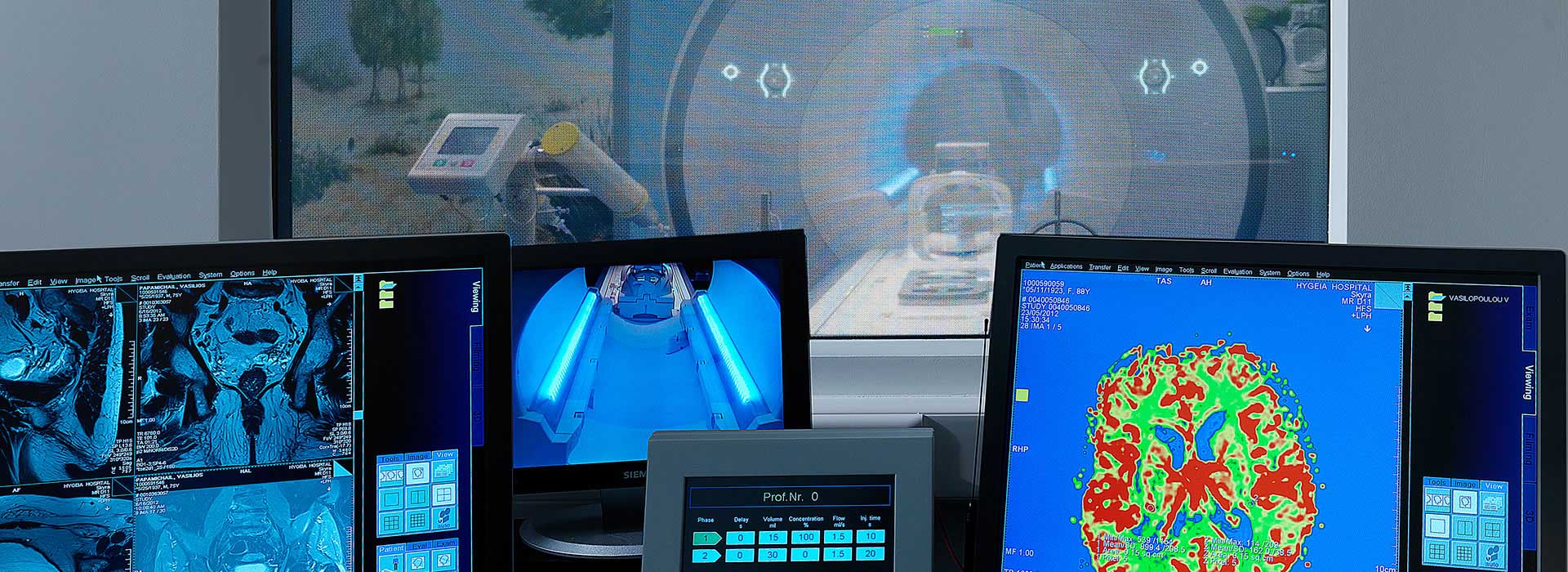
At HYGEIA Hospital, the department of research and development of Cardiac MR was developed in July 2002. The department uses a two 1.5 Tesla MR scanners (Philips Intera and Achieva) with up to date software that can support all cardiac examinations. The examinations are stored in a PACS system, from which they can be readily retrieved upon request. Clinical examinations are performed on a daily basis for clinical indications. The clinical experience from the first 1000 cases has been published in the medical press. The HYGEIA Cardiac MR Center actively participates in clinical research protocols and has developed scientific collaborations with recognized institutions in Greece and abroad.
- Cardiac Magnetic Resonance Imaging can accurately depict myocardium anatomy and of the great thoracic vessels. Apparent clinical applications include: evaluation of the aorta for dissections and aneurysms, assessment of pericardial thickness and constrictive pericarditis, and imaging of cardiac and pericardial tumors.
- Cardiac Magnetic Resonance Imaging is particularly useful for the evaluation of anatomy in various forms of cardiomyopathy, including: acute myocarditis, dilated and hypertrophic cardiomyopathies and in various forms of infiltrative cardiomyopathies (sarcoidosis, amyloidosis, hemosiderosis, etc). Specifically for patients undergoing multiple blood transfusions, cardiac magnetic resonance imaging has been proposed as the most reliable method to follow-up myocardial signal changes attributed to tissue iron deposition, and guide chelation therapy.
- In arrhythmogenic right ventricular dysplasia, Cardiac Magnetic Resonance Imaging is the only non-invasive modality that can visualize myocardial fat infiltration, along with the associated regional wall motion and thickening abnormalities.
- Cardiac Magnetic Resonance Imaging is the most accurate method for left and right ventricular function assessment. For functional studies, “cine” sequences are typically obtained in synch with the electrocardiographic signal. Stacks of multi-phase images are typically obtained during short breath-hold periods.
- The quantitative evaluation of blood flow is possible with a technique called “phase contrast” or “velocity-encoding”. This method can accurately measure flow in any direction and can be used for quantitatively measuring cardiac output, valve insufficiencies, as well as in patients with left-to-right shunts.
- As Cardiac Magnetic Resonance Imaging can provide a reliable and thorough evaluation of cardiac anatomy and function, this method has particular advantage for evaluating children and adults with congenital heart disease. At HYGEIA Hospital examinations are carried out even in newborns and babies, with appropriate anesthesiological support.
- One of Cardiac Magnetic Resonance Imaging applications that was quickly established in clinical practice, is the detection of myocardial viability.
- In patients who have suffered a myocardial infarction, Cardiac Magnetic Resonance Imaging demonstrates accurately the extent of the infarction, whether it is subendocardial or transmural, by administering of paramagnetic contrast medium (gadolinium).
Magnetic Resonance Coronary Angiography has always been considered as the most attractive application of Cardiac Magnetic Resonance Imaging.
In recent years, the origin and proximal segments of the coronary arteries can be reliably visualized in the majority of patients. This method has already been established as “the method of choice in the assessment of patients with coronary artery anomalies”.
The clinical role of Magnetic resonance Coronary Angiography for diagnosing coronary artery disease is limited to specialized applications (e.g. the exclusion of proximal three-vessel disease), and should be performed with caution in highly selected patients and only in specialized centers with experience in this technique, such as the cardiac MRI Department of HYGEIA Hospital.
HYGEIA Hospital has been one of 17 hospitals worldwide which participated in a large international multicenter research clinical trial of predicting myocardial function improvement, through viability assessment in patients with ischemic cardiomyopathy and severe cardiac dysfunction undergoing bypass surgery or angioplasty.
Educational seminars are been offered annually at HYGEIA. These programs target Cardiologists or Radiologists who have interest and seek training in CMR. One to three trainees will participate in each program. Selection will be on a first-come first-served basis until all available positions are covered. The maximal numbered of trainees will be strictly enforced. The language of the seminar will be English (or Greek if only Greeks participate).
Educational goals:
- The understanding of
- The basic physics principles of MR imaging
- The basic imaging sequenses
- How to organize a CMR examination
- The finding at various disease entities
- The clinical utility of CMR
- The advantages and challenges of CMR compared to other modalities of cardiac imaging.
During 2015 the program took place from 2 – 13 Νovember. During this period there were daily lectures on cardiac MR, observation of the regular clinical examinations performed at the department, demonstration of didactic cases and discussion, voluntary involvement with research protocols and interaction and discussion with the training staff.
The trainee’s received a syllabus with educational material and a certificate of participation.The Scientific head of the course is Dr. Peter G. Danias
 Contact Us
6th floor
Οffice 6.1
(Erythrou Stavrou 5 – opposite Hygeia Hospital)Telephone
+30 210 686 7660
Email
CardiacMRI@hygeia.grMedical Team
- Head of Department
-
 Danias Petros
 - Αttending Physicians
-
 Andreou Ioannis
 -
 Efthimiadou Roxani
 -
 Filippi Vasiliki
 -
 Karatzis Emmanouel
 -
 Kritikos Nikolaos
 -
 Mourmouris Christos
 -
 Rousakis Arkadios

- © 2007-2024 HYGEIA S.M.S.A.

- Services