























OPTOVUE VISIONIX SOLIX OPTICAL COHERENCE TOMOGRAPHY ANGIOGRAPHY (OCTA)
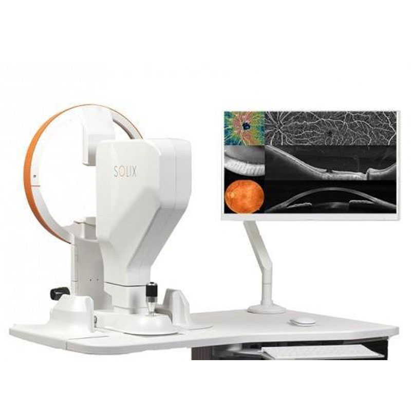
SOLIX is a new technology built upon a proven foundation of high-speed Spectral Domain OCT. This FullRange platform empowers practitioners to identify and manage numerous pathologies from the front of the eye to the back for a vastly superior diagnostic experience.
Visionix Optical coherence tomography angiography (OCTA) is a noninvasive imaging modality used to diagnose and manage retinal and choroidal vascular disease.OCTA has the ability to detect subtle lesions that are not identifiable during clinical examinations. OCTA has a role in understanding tumour vascularity.
SOLIX delivers multiple tools for a new generation of disease management that improve throughput and enable superior patient care:
- FullRange anterior segment imaging to capture the entire anterior chamber in a single scan
- External IR imaging to enable evaluation of Meibomian glands of the upper and lower lids without a dedicated imaging device
- Proven glaucoma analytics that combine structural and vascular images and measurements • FullRange retinal imaging that allows wide and deep imaging of the retina, choroid and vitreous…even in highly myopic patients
- Optovue’s industry-leading AngioVue® OCT Angiography (OCTA) for non-invasive 3D visualization and quantification of retinal vasculature*
- Fundus and external color photography
- Wellness capabilities that have become part of a new standard of care for patients suspected of both retinal pathologies and glaucoma
- Ultra-fast spectral-domain technology produces a wide and deep field of view that does not compromise image resolution
- Multi-volume merge averages four scan volumes to deliver high-density images with pristine clarity
- 3D vessel rendering enables real-life visualization of retinal vasculature and vascular connectivity • 3D PAR 2.0 rapidly removes the majority of projection artifact from the deep plexus to simplify image interpretation and produce more reliable quantification
- New segmentation algorithms dramatically improve Bruch’s membrane and RPE segmentation for more confident assessment – even in highly diseased eyes
- DualTrac™ Motion Correction Technology with enhanced visualization combines real-time tracking and patented postprocessing to enable true 3D correction of distortion in all directions for ultra-precise motion correction.
Available at the Ocular Oncology Center Pediatric and Adult of HYGEIA.
Visit the Center