

























OPTOVUE VISIONIX SOLIX ANTERIOR SEGMENT OPTICAL COHERENCE TOMOGRAPHY (AS-OCT)
SOLIX is a new technology built upon a proven foundation of high-speed Spectral Domain OCT. This FullRange platform empowers practitioners to identify and manage numerous pathologies from the front of the eye to the back for a vastly superior diagnostic experience.
OCT provides detailed images of ocular tissue structure, enabling early detection of anomalies and monitoring of ocular disease progression.
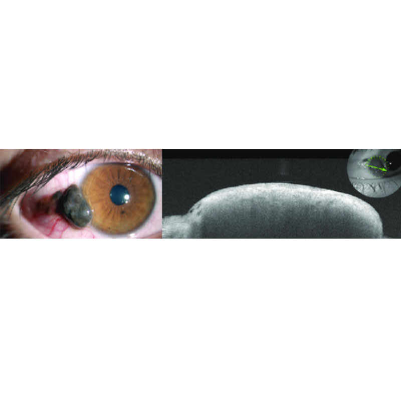
AS-OCT has proved to be an indispensable diagnostic tool in diagnosing and managing conjunctival and corneal pathologies.
AS-OCT has been widely used in the diagnosis of ocular surface tumours providing in vitro “optical biopsy” features that offers similar if not better results in demonstrating internal tumor features.
SOLIX delivers multiple tools for a new generation of disease management that improve throughput and enable superior patient care:
- FullRange anterior segment imaging to capture the entire anterior chamber in a single scan
- External IR imaging to enable evaluation of Meibomian glands of the upper and lower lids without a dedicated imaging device
- Proven glaucoma analytics that combine structural and vascular images and measurements • FullRange retinal imaging that allows wide and deep imaging of the retina, choroid and vitreous…even in highly myopic patients
- Fundus and external color photography
- Wellness capabilities that have become part of a new standard of care for patients suspected of both retinal pathologies and glaucoma
- Ultra-fast spectral-domain technology produces a wide and deep field of view that does not compromise image resolution
- Multi-volume merge averages four scan volumes to deliver high-density images with pristine clarity
- 3D vessel rendering enables real-life visualization of retinal vasculature and vascular connectivity • 3D PAR 2.0 rapidly removes the majority of projection artifact from the deep plexus to simplify image interpretation and produce more reliable quantification
- New segmentation algorithms dramatically improve Bruch’s membrane and RPE segmentation for more confident assessment – even in highly diseased eyes
- DualTrac™ Motion Correction Technology with enhanced visualization combines real-time tracking and patented postprocessing to enable true 3D correction of distortion in all directions for ultra-precise motion correction.
Available at the Ocular Oncology Center Pediatric and Adult of HYGEIA.
Visit the Center