


- HYGEIA
- Virtual tour
- Services
- Maternity Clinic
- Maternity Clinic Medical Services
- Paediatric Clinic
- Paediatric Clinic Medical Services


- HYGEIA
HYGEIA
- Virtual tour
Virtual tour
- Services
Services
- Maternity Clinic
Maternity Clinic
- Maternity Clinic Medical Services
Maternity Clinic Medical Services
Echocardiography
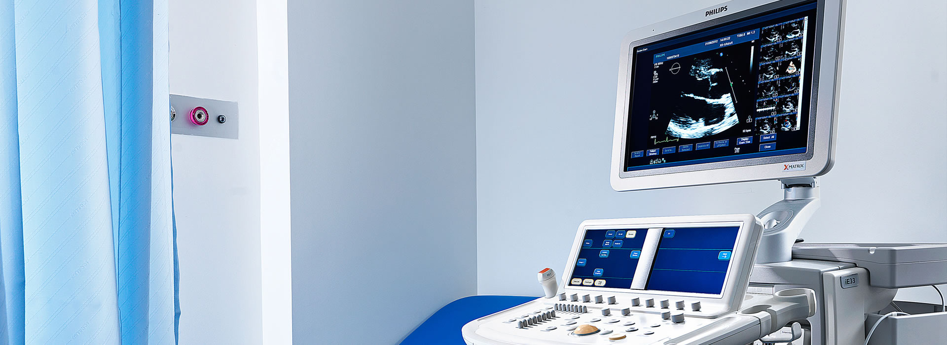
The Cardiac Ultrasound Division is a laboratory that is fully-equipped with state-of-the-art devices and two- and three-dimensional imaging capabilities. It is able to meet the needs of inpatients and outpatients.
The following tests are performed:
- Two-dimensional and three-dimensional transthoracic echocardiography (2D, 3D, echo, colour Doppler).
- Transoesophageal echocardiography (2D & 3D).
- Intraoperative transoesophageal echocardiography during cardiac surgery and intraprocedurally during MitraClip and transcatheter aortic valve implantation (TAVI).
- Dynamic echocardiography (stress echo), with or without the use of a contrast agent.
- Echocardiography that features tracking algorithms for assessing global longitudinal strain. This is the appropriate method for monitoring cardiac function in cancer patients on chemotherapy.
Our imaging
This method is based on high-frequency sound waves (i.e. sounds that cannot be perceived by the human ear) that bounce back to the transducer (the device that the doctor slides across your chest) and are converted into images using computers. Therefore, by sending an ultrasound beam to the lateral chest wall, we can examine the heart as it beats. In doing so, we can study and accurately measure the size of the cardiac chambers, while being able to assess the contraction of the heart chambers and the function of the heart valves.
Triplex – colour cardiac ultrasound
This is one of the most comprehensive non-invasive tests of the heart and is used to evaluate heart valve function. Apart from the morphological information that simple cardiac ultrasound provides, the addition of colour allows us to record the velocity with which blood flows through the valves, and specific calculations allow us to understand whether they are working properly
Stress Echo
This is a dynamic test that has been established over the last 15 years as an alternative non-invasive technique for the diagnosis of coronary heart disease, but also for the assessment of coronary heart disease patients. This is a special type of ultrasound that involves the intravenous administration of a drug that “tires” the heart. During this procedure, we try to visualise whether the force with which the heart muscle contracts is affected, thereby indirectly concluding whether there is any blockage in the coronary arteries.
Transoesophageal ultrasound
This test is performed in a manner that is similar to a gastroscopy. By inserting the transducer through the mouth and lowering it down the oesophagus, we come very close to the heart and are therefore able to visualise the heart chambers and valves in great detail. This method can be used when access via the chest wall does not provide satisfactory imaging. It is used in 5-10% of patients who require echocardiographic assessment.
Intraoperative – transoesophageal echocardiography
This test allows patients to be monitored during cardiac surgery procedures, resulting in a more accurate assessment of the patient’s condition (preoperatively), haemodynamic status (intraoperatively) and procedure results (immediately after surgery). Most significantly, this method contributes to the prompt repair of residual lesions that are discovered immediately after surgery.
3D heart ultrasound
From a practical aspect, this is a three-dimensional image of the heart. It enables the anatomy of the heart to be studied with greater accuracy and detail. This method is extremely useful in the case of valvulopathies and congenital diseases, where the surgeon can acquire information about the type and method of surgery with greater accuracy and detail.
The Heart Ultrasound Division is equipped with portable devices that allow emergency echocardiography to be performed in the Intensive Care Unit and Haemodynamic Laboratory, as well as on patients who cannot be moved from their rooms.
 Contact Us
210 686 7210
Medical Team
- Director
-
 Levantakis Ioannis
 - Associate Director
-
 Gatos Spyridon
 - Collaborators
-
 Fountoulakis Petros
 -
Kapelakis Ioannis

- Services
- Virtual tour
- HYGEIA
- Paediatric Clinic
- Services
- Virtual tour