


- HYGEIA
- Virtual tour
- Services
- Maternity Clinic
- Maternity Clinic Medical Services
- Paediatric Clinic
- Paediatric Clinic Medical Services


- HYGEIA
HYGEIA
- Virtual tour
Virtual tour
- Services
Services
- Maternity Clinic
Maternity Clinic
- Maternity Clinic Medical Services
Maternity Clinic Medical Services
PET / CT
The HYGEIA Hospital PET/CT Department was the first to operate in Greece, in 2004, and is certified to ISO 9001:2008. The PET/CT scan is designed and performed by a team of specialized scientists (nuclear physicians, radiologists, radiophysicists and technologists), who have been trained accordingly in major healthcare centers abroad.
The main PET applications pertain to oncology and neurology, with cardiology close behind. PET/CT contributes to accurate diagnosis, staging and restaging of cancer patients. It has proven significant in managing patients with neoplasms and selecting the most suitable treatment.
Lately, the PET/CT findings are used by the Radiation and Oncology Center, for optimal radiotherapy planning, so that patients achieve the best possible treatment results, with the least possible radiation burden to healthy tissues.
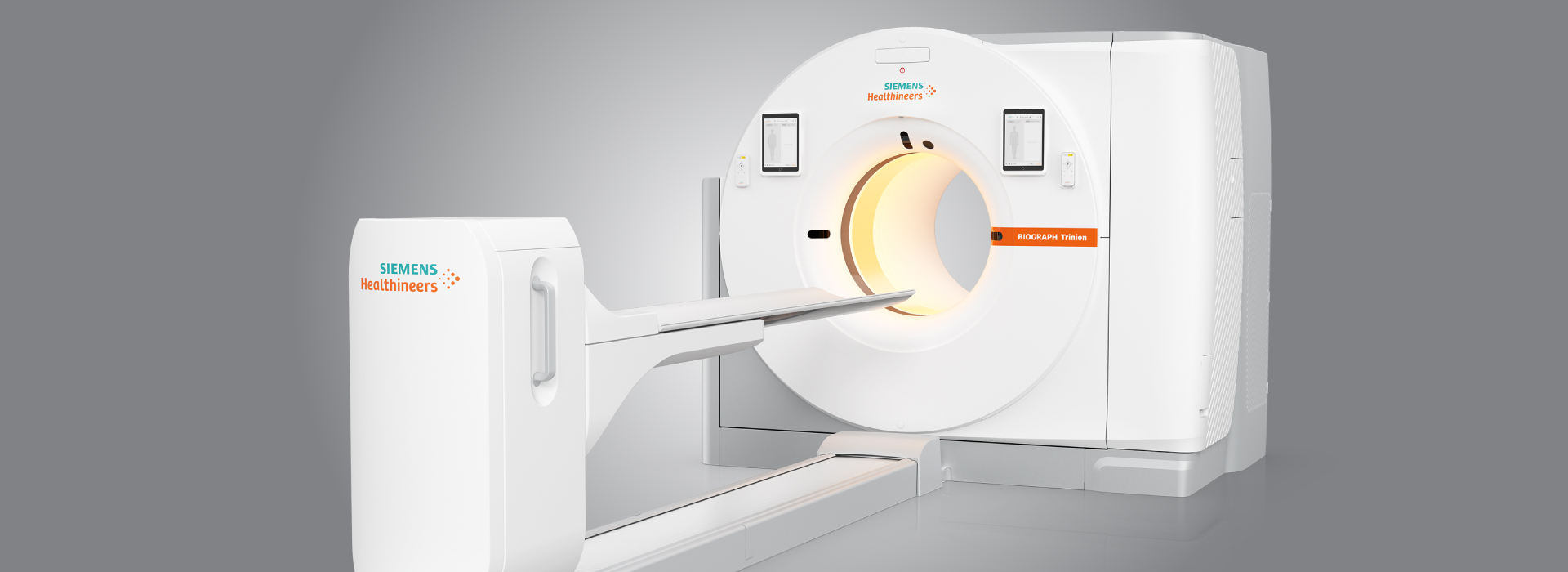
In August 2012, the PET/CT Department was equipped with the Siemens Biograph 16 PET/CT scanner. This scanner shows detailed anatomy and biological processes at the molecular level, with just one non-invasive procedure. The Siemens Biograph 16 features LSO detector design, exceptional imaging quality, 16-slice CT detectors and the SYNGO multimodality computer system.
What is a PET/CT scan?
It is an imaging test that combines two techniques, Positron Emission Tomography (PET), a cutting-edge Nuclear Medicine imaging technique, and Computed Tomography (CT) in a single system. The device is made up of a multi-slice helical CT scanner and a latest generation positron scanner with LSO crystals, which create images faster, thus offering quicker and more accurate visual representation of the patient. It combines two techniques in one exam, so that patients do need to move between machines for CT images, which depict the anatomy of the organs, and PET images, which depict the function and metabolism of said organs.The PET/CT scanner software (SYNGO) reconstructs and combines the PET and CT images. By fusing the body’s anatomical and metabolic images, it visually records living tissue, both healthy and pathological; this also justifies its name: BIOGRAPH (biological recorder).
Where is the PET/CT scanner used?
• On oncology patients: For initial diagnosis of the disease, staging, fast assessment of the treatment result (faster than conventional methods) and detection of recurrence. The method may prove useful is detecting an unknown primary neoplasm when the rest of the imaging techniques are unable to diagnose this, while it has contributed to designing radiotherapy treatment plans for patients about to undergo radiotherapy.
• On neurology patients: For early diagnosis of Alzheimer’s, differential diagnosis of various types of dementias and detection of epileptic foci in epileptic patients.
• On cardiology patients: For diagnosis of myocardial ischemia and detection of viable myocardium in selected patients when the attending physician believes the other diagnostic screening tests are not as effective.Why is the introduction of the PET/CT method so important?
• It achieves earlier and more accurate diagnosis of the disease, and consequently, faster and more effective treatment.
• It achieves significant savings in patient management, which is important to the National Health System and insurance companies. Note that the application of PET/CT scans changed the manner in which oncology patients were being treated at a rate of 30-35%; i.e. in a third of cancer patients, the treatment that had been planned prior to the PET/CT changed after the imaging results produced by this method. This percentage is significant, translating into major financial benefits for a health system.• Whole-Body PET/CT (from skull base to mid thigh)
• Brain PET/CT (from top to base of the skull)
• Lower Limb PET/CT (from mid thigh to feet)
• Whole-Body and Brain PET/CT
• Whole-Body and Lower Limb PET/CT
• Whole-Body, Brain and Lower Limb PET/CTImaging radiopharmaceuticals
• 18F-FDG
• 18F-FCH (fluorocholine) (mainly for the imaging of prostate cancer and brain lesions)
• 18F-FLT (fluorothymidine) (mainly for brain lesions)Results
The test results are available 2 working days after the exam.Read the instructions about the PET/CT scan
Read the instructions on Radiation Protection for patients about undergo a PET/CT scan
 Contact Us
Medical Team
- Director of Imaging Departments of Hygeia Hospital
-
 Andreou Ioannis
 - Directors
-
 Efthimiadou Roxani
 -
 Pipikos Theodoros
 - Assistant Director
-
 Vogiatzis Merkourios
 - Nuclear Physicians
-
 Doika Efstathia
 -
Ilia Eliverta
- Radiologists
-
 Filippi Vasiliki
 -
Harokopakis Aggelos
-
Hopkins - Hatzopoulou Anabell
-
Kommata Spyridoula
-
 Laspas Fotis
 - Radiophysicists
-
 Dalianis Konstantinos
 -
 Gogos Konstantinos

- Services
- Virtual tour
- HYGEIA
- Paediatric Clinic
- Services
- Virtual tour